
Robotic
Obturator Nerve
Decompression
Beyond Groin Pain—Identifying the True Source
Obturator nerve–related pain is frequently underdiagnosed and often mistaken for musculoskeletal or orthopedic conditions. Many patients undergo prolonged treatment for hip, groin, or adductor-related pain without resolution, as the true source lies within the deep pelvic nerve anatomy.
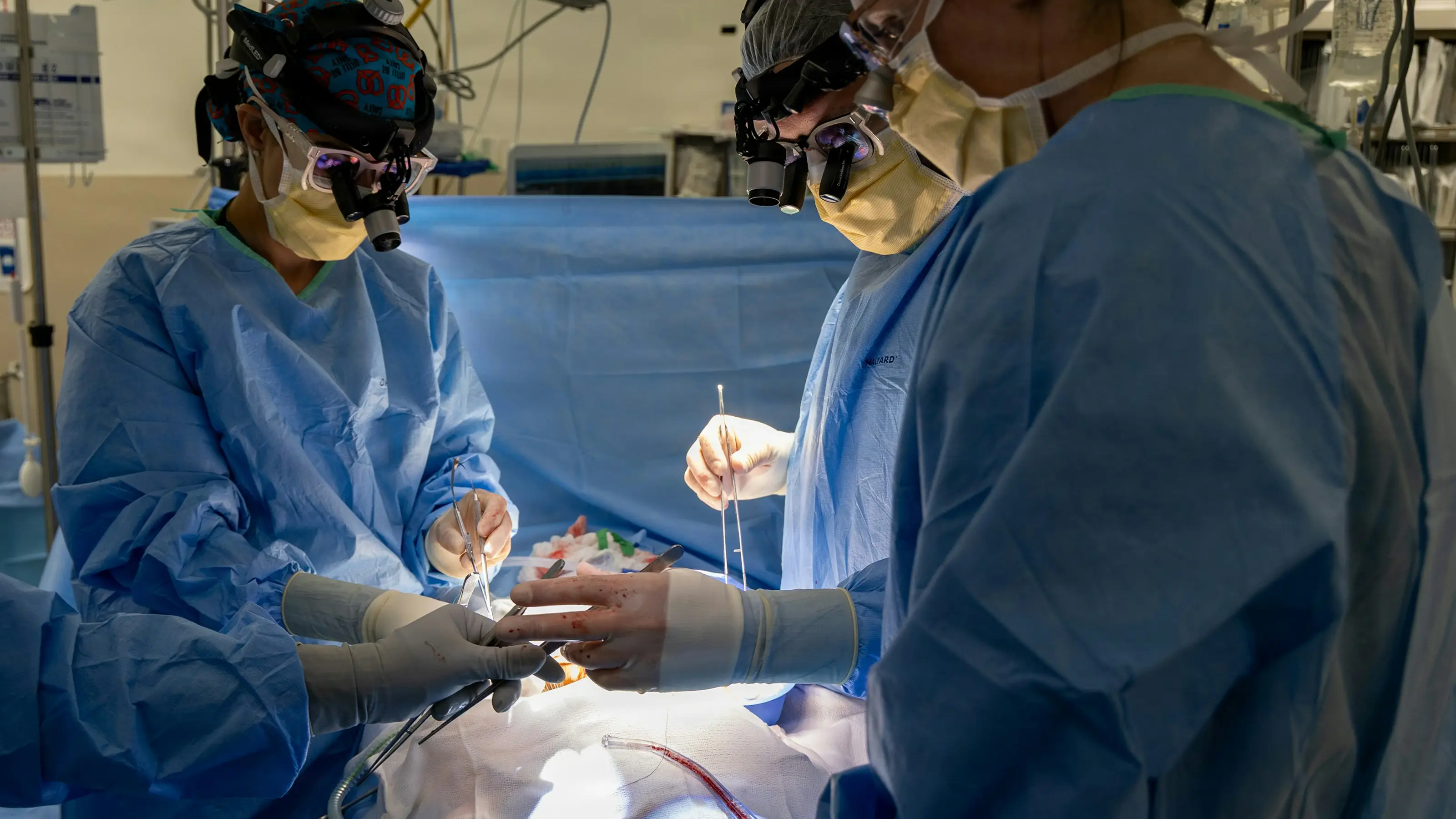
At our center, we specialize in robotic-assisted intrapelvic obturator nerve decompression, allowing precise identification and treatment of pathology affecting the nerve at its origin and along its course through the pelvis.
What Is Obturator Nerve Entrapment?

The obturator nerve arises from the lumbosacral plexus and travels along the pelvic sidewall into the deep obturator space, eventually exiting through the obturator canal to supply the medial thigh.
Along this course, the nerve may be affected by:
- Inflammatory adhesions
- Fibrosis and scarring
- Deep infiltrating endometriosis
- Compression within the obturator canal or surrounding fascia
Because much of this pathology occurs deep within the pelvis, it is often not visible through standard imaging or accessible through external approaches.
Symptoms
Patients with obturator nerve involvement may experience:
- Medial thigh pain or burning sensation
- Groin pain radiating to the inner thigh
- Pain with leg adduction or walking
- Weakness or instability of the adductor muscles
- Deep pelvic or paravaginal discomfort
- Overlap with pelvic pain syndromes
Symptoms are often persistent and misdiagnosed as hip or musculoskeletal pathology.
Why This Happens
Common causes include:
- Pelvic neuroinflammation
- Endometriosis involving the pelvic sidewall or obturator space
- Inflammatory adipose tissue encasing the nerve
- Post-surgical fibrosis or chronic scarring
- Compression along the fascial planes of the pelvic sidewall
In many cases, obturator nerve involvement is part of a broader multinodal pelvic nerve condition.
Why Standard Treatments Often Fail
Traditional treatments may include:
- Orthopedic interventions
- Physical therapy
- Pain medications or injections
These approaches often fail because they do not address intrapelvic nerve pathology.
External surgical approaches to the obturator nerve are limited by:
- Restricted visualization
- Inability to assess the nerve at its origin
- Limited access to surrounding pelvic structures
Our Surgical Approach
We utilize a robotic intrapelvic approach, allowing direct access to the obturator nerve along its entire pelvic course and providing:
- Safe development of the retroperitoneal space
- Precise identification of the obturator nerve at its origin
- Direct access to the deep obturator space and obturator canal
- High-definition visualization for targeted decompression
Comprehensive Access to the
Obturator and Pelvic Sidewall Region
A key advantage of this approach is the ability to achieve direct access and exceptional visualization of the deep obturator space and entire pelvic sidewall.
This allows the surgeon to:
- Trace the full trajectory of the obturator nerve from its origin
- Identify and release fibrotic bands, inflammatory adhesions, or nodular disease
- Perform precise neurolysis within confined anatomical spaces
Importantly, this approach enables evaluation and treatment of adjacent neural structures that may contribute to overlapping symptoms, including:
- Lumbosacral plexus branches
- Pudendal nerve
- Sciatic nerve (proximal segment)
- Posterior femoral cutaneous nerve
In addition, the surgeon gains enhanced visualization of surrounding pelvic compartments, including:
- Paravaginal and paraurethral spaces
- Endopelvic fascia and levator ani origin
- Pelvic neurovascular structures along the sidewall

A Truly Comprehensive Neuroanatomical Approach
Unlike traditional approaches that focus on a limited segment of the nerve, this technique allows for:
What to Expect
Minimally invasive robotic procedure
High precision with minimal blood loss
Typically outpatient or short hospital stay
Surgical time varies depending on disease complexity
Recovery & Nerve Healing

Nerve recovery is gradual and individualized:
- Early symptom improvement may begin within weeks
- Continued healing occurs over several months
- Adjunct therapies may be used to support nerve recovery
Due to the minimally invasive nature of the approach, patients typically experience:
- Less postoperative pain
- Faster recovery compared to traditional approaches
Who is a candidate?
You may be a candidate if you have:
- Chronic groin or medial thigh pain
- Pain worsened by walking or adduction
- Persistent symptoms despite conservative care
- Suspected or confirmed pelvic endometriosis
- History of pelvic surgery with ongoing neuropathic symptoms
The Key Difference
Traditional approaches attempt to treat symptoms at the periphery.
Our approach allows us to access the nerve at its origin, identify the cause, and treat the entire pelvic nerve environment.